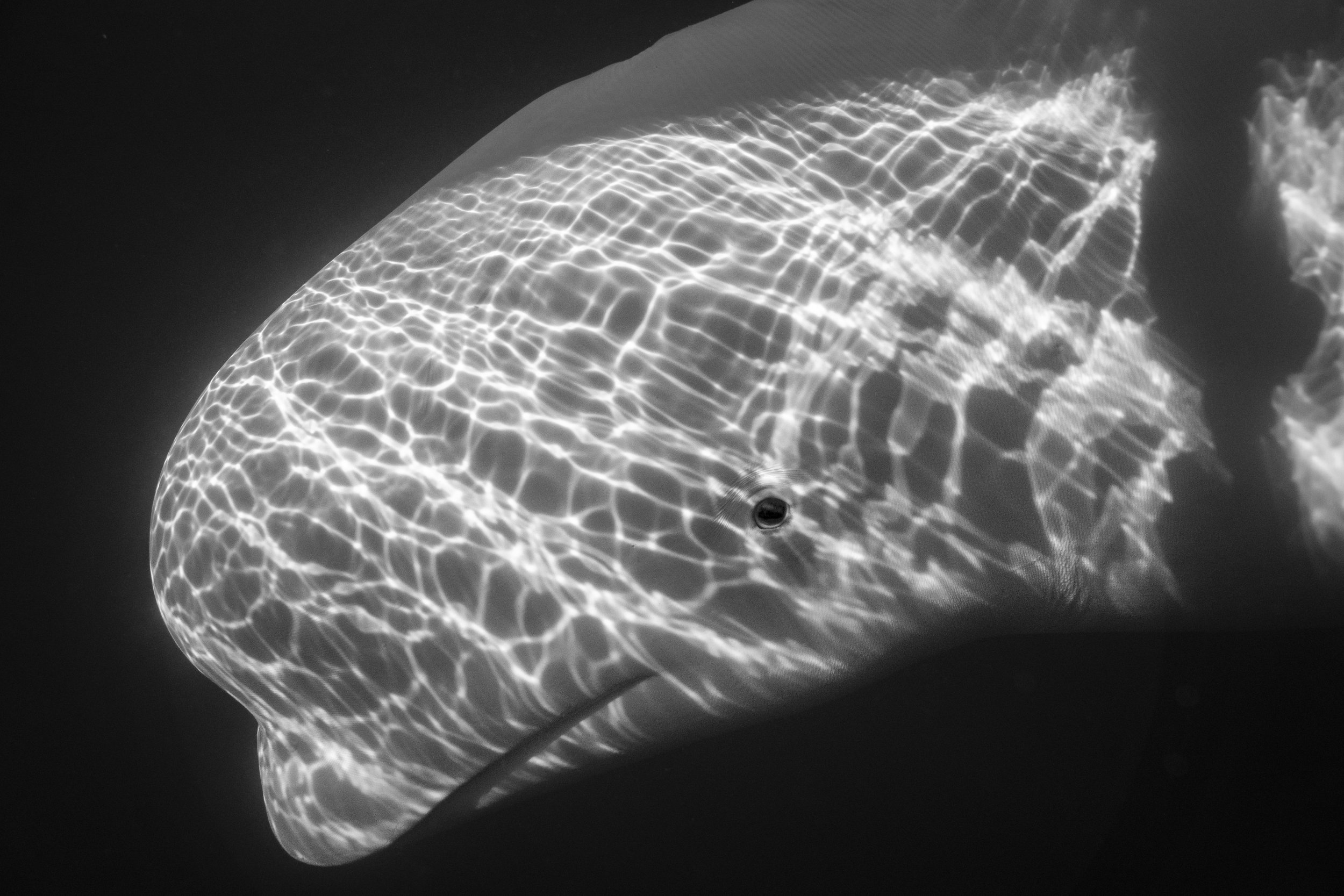
Hvaldimir’s Full Autopsy Report

Sør-Vest Police District
Lagårdsveien 6,
4010 Stavanger
Reference: 2024-40-108/P80
Date: 23.09.2024
Case History:
According to Kathrine Ryeng from the Institute of Marine Research in Tromsø, Hvaldimir, the beluga whale, had appeared healthy and in good condition until recently. The whale was found dead, floating in the water, on the afternoon of Saturday, August 31, 2024. A preliminary report was sent on September 6, 2024.
Autopsy Report:
Received on September 2, 2024: The carcass of a beluga whale, male, adult. Age estimated at 14-16 years, weight approximately 1000 kg based on external examination, and length measured at 4.23 meters.
The carcass was in an advanced state of decomposition, which complicated the autopsy, assessment, and interpretation of findings.
The whale was in good nutritional condition with thick layers of blubber (3-9 cm depending on location). However, the stomachs and the front parts of the small intestine were mostly empty.
During handling, the skin over large parts of the whale's right side and head detached (these were the parts lying against the ground during storage before the autopsy).
Findings:
- Seven superficial skin wounds were found in the area of the posterior chest, abdomen, and near the tail fin, with varying depths (from 2 to 10 mm). The wound edges were jagged and appeared to have been caused by birds pecking through the skin.
- On the underside of the chest, between the front flippers and slightly to the left of the midline, there were two perforations in the skin, 19.5 cm apart and 0.5 cm in diameter.
- The first perforation channel extended through the skin and the entire blubber layer (7 cm long). The surrounding tissue was not bloody or torn.
- The second perforation was shorter, less than 1 cm long, and did not penetrate the blubber layer. Both holes had dark wound edges surrounded by circular impressions.
No metal fragments were found on X-rays of the chest and head.
A 35 cm long wooden stick was found lodged in the whale’s mouth, partially embedded in the soft tissue between the teeth/jawbone and the base of the tongue, extending towards the throat. The front end of the stick was pressed against the hard palate, causing the mucous membrane to detach. The embedded end of the stick had a rounded tip, while the exposed end was splintered. The embedded part of the stick was red in color. The stick was partially covered in bark and showed no signs of recent human modification.
Internal Findings:
- More than 5 liters of red-black, blood-tinged fluid were found in the chest cavity, and over 10 liters in the abdominal cavity.
- The organs in both cavities had significantly degraded and were darkly discolored.
- Ribs 3 and 4 on the right side were broken near the breastbone but were not significantly displaced.
**Microscopic (Histological) Examination:**
- Samples from the spleen, kidney, lung, skin, gastric and back muscles, blubber, and the soft tissue around the stick in the mouth showed advanced decomposition.
- The tissue around the stick contained bleeding, bacterial clusters, plant material, and abundant large cells interpreted as macrophages, typical of a foreign body reaction (granulomatous reaction).
- In one of the bird-inflicted wounds, tissue degradation and bacterial clusters were found but with no inflammation reaction.
Microbiology:
- A mixed bacterial flora (more than three different species) was found in muscle and lung tissue samples.
- Several bacteria, including *Proteus mirabilis* and *Clostridium septicum*, were abundant in the stomach contents and feces.
- *Campylobacter sp.*, *Salmonella sp.*, and other pathogens were not detected in the samples.
Parasitology:
- Anisakid parasites were found in the stomach contents, with 100 eggs per gram detected in fecal samples, indicating a mild parasitic infection.
Virology:
- Tests for Influenza A virus were negative in the throat, trachea, and lung samples.
Diagnosis:
- Foreign body in the mouth with granulomatous infection.
- Advanced decomposition (cadaverosis).
- Mild parasitic infection.
Conclusion:
Due to the advanced state of decomposition, the findings must be interpreted cautiously. However, there is no evidence to suggest that the whale died as a result of being shot. The described wounds are not considered fatal. The stick lodged in the mouth, on the other hand, must have caused significant pain and discomfort and likely made it difficult for the whale to eat. Furthermore, the wound may have served as an entry point for bacteria, leading to an infection and/or toxin production, potentially causing the whale’s death through sepsis (blood poisoning) and/or toxemia (poisoning).
Veterinarians:
Michaela Falk, PhD,
Bjørnar Ytrehus, Dr. med. vet., Specialist in wildlife health
